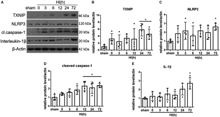
FIGURE 1.

Temporal expression of endogenous TXNIP, NLRP3, cleaved caspase‐1 and interleukin‐1β in the ipsilateral brain hemisphere after hypoxia‐ischemia (HI). A, Representative pictures of Western blot data. B, Western blot data analysis showed that endogenous TXNIP expression levels significantly increased at 24 and 72 h post‐HI. C, Western blot data showed that endogenous NLRP3 expression levels increased, reaching significance at 72 h after HI. D, Cleaved caspase‐1 expression was significantly increased at 12, 24 and 72 h post‐HI. E, Active interleukin‐1β expression was increased after HI, reaching statistical significance at 72 h after HI. *P < 0.05 vs sham. Data are represented as mean ± SD, n = 4 for each group
