FIGURE 4.

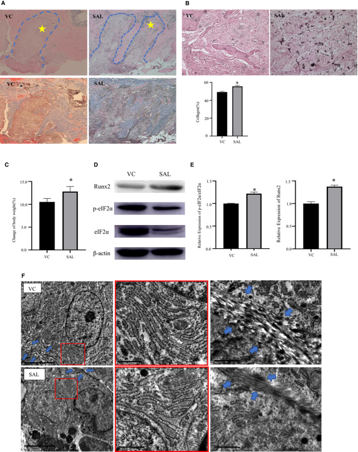
Suppressing ER stress could protect osteogenesis in vivo. A, Representative photomicrographs of tooth extraction sockets in the salubrinal (SAL) and vehicle control (VC) groups. The blue dotted line indicates the tooth extraction socket, and the star indicates irregular woven bone. SAL group significantly increased the number of collagen fibres. Top: HE staining, 50×; bottom: Masson's tricolour staining, 50×, blue fibres in connective tissue represent collagen. B, Representative photomicrographs of ALP staining (200×) in tooth extraction sockets in the SAL and control groups. C, The percentage change in body weight. Body weight increased after using salubrinal. D‐E, Representative Western blots of the protein expression levels of eIF2α, p‐eIF2α and Runx2 in the bone tissue around the tooth extraction wounds with different drug interventions. p‐eIF2α/eIF2α and Runx2/β‐actin protein expression was higher after salubrinal use. F, Transmission electron microscope images showing the ultrastructural changes of the rough ER (red square) and the formation of collagen fibres (blue arrows) in osteoblast around the sockets in the SAL and control groups (top: PBS, bottom: SAL) Left scale bar = 5 μm, middle and right scale bar = 1 μm. Data were presented as the ratio with β‐actin and mean ± SD. The asterisks (*) represent P < .05(n = 9)
