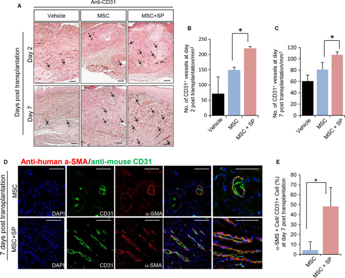
Figure 5.

SP improves MSC‐induced angiogenesis at the wound site. Histological analysis of wounded tissue at day 7 post‐wound was performed. (A) Histological analysis of the vasculature was performed via staining for CD31 (B‐C) Quantification of CD31 (+) vasculature was performed at days 2 and 7 post‐transplantation. (D) Immunofluorescence staining of skin tissues with anti‐human α‐SMA and anti‐mouse CD31 to detect the localization of transplanted MSCs at the host vasculature. (E) Transplanted human α‐SMA (+) MSCs‐encircled vessels of total mouse CD31 (+) vessels were quantified. α‐SMA (+) human MSCs was shown in red and CD31 (+) mouse vascular endothelial cells are shown in green. Values of P < .05 were considered statistically significant (*P < .05). The data are represented as mean ± SD of three independent experiments. N = 8 for each group. Scale bar: 100 μm
