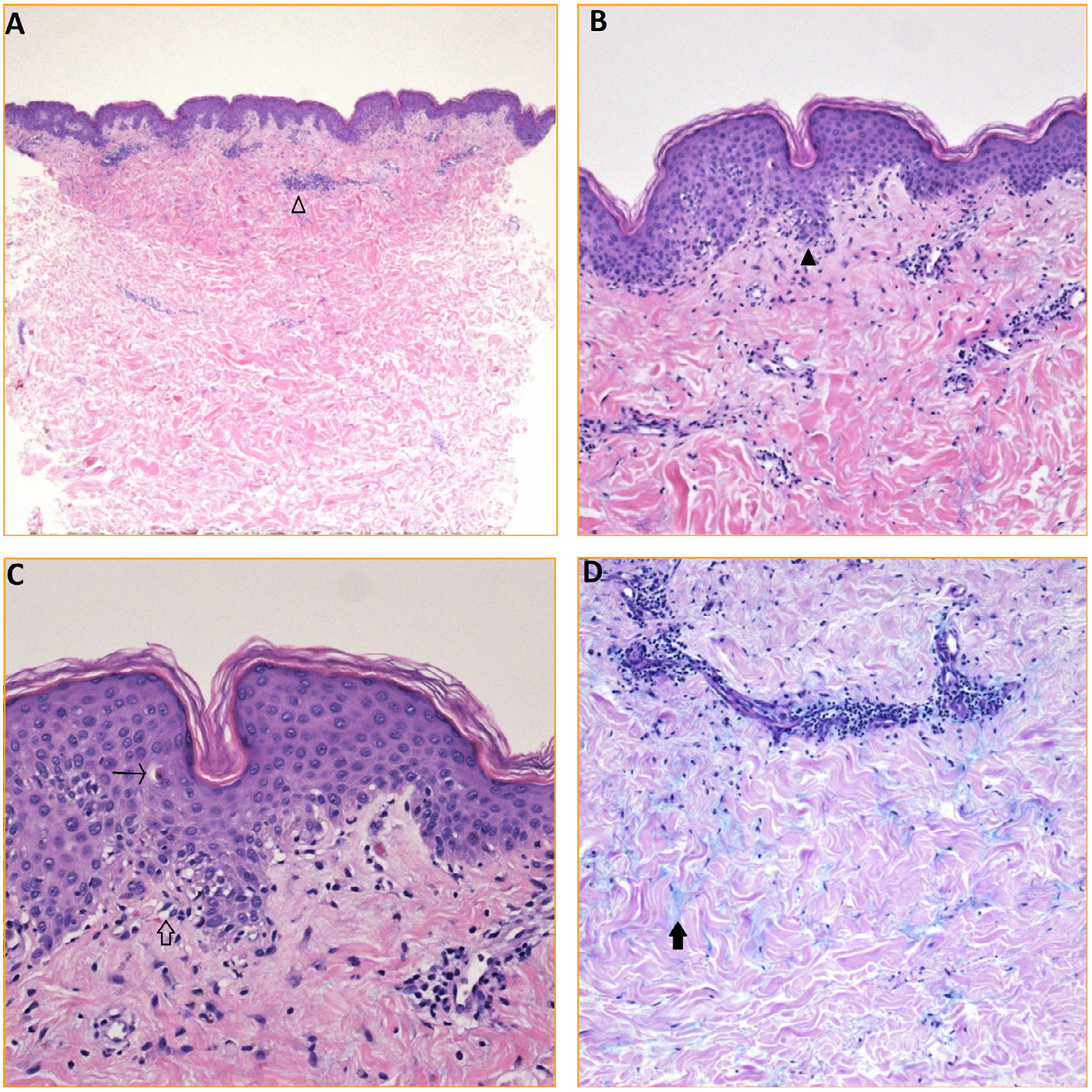
FIGURE 2.

Skin biopsy findings. (A) H&E (20×): punch biopsy of skin shows a superficial perivascular infiltrate of entirely lymphocytes (empty arrowhead). (B) H&E (100×): subtle interface change characterized by vacuolar alteration along the dermo-epidermal junction (black arrowhead). (C) H&E (200×): obvious vacuolization along the dermo-epidermal junction (empty arrow) with rare individually necrotic keratinocytes (arrow). (D) Alcian blue (100×): copious mucin (black arrow) situated between bundles of collagen within the reticular dermis.
