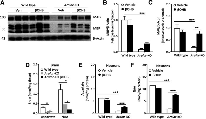
Figure 6.
Effects of βOHB on cortical myelin protein levels and Asp/NAA brain and primary cortical neurons content. A, Representative Western Blotting images of MBP and MAG, with their respective densitometric histograms (B, C) in WT and aralar-KO brain cortex after 290 mg/kg/d βOHB intraperitoneal injections. β-Actin used as charge control. Mean ± SEM (n = 5–7 mice per group); ***p ≤ 0.001, **p ≤ 0.01 (one-way ANOVA followed by Newman–Keuls multiple comparisons t test). D, Asp and NAA levels after 290 mg/kg/d βOHB intraperitoneal injections in WT and aralar-KO brain. Mean ± SEM (n = 3 mice per group); **p ≤ 0.01, *p ≤ 0.05 (one-way ANOVA followed by Newman–Keuls multiple comparisons t test). E, F, Asp (E) and NAA (F) levels after 5 mm βOHB 96-h treatment in WT and aralar-KO primary neuronal cultures. Mean ± SEM from two to four embryos per condition measured in duplicates; ***p ≤ 0.001 (one-way ANOVA followed by Newman–Keuls multiple comparisons t test).

