
Figure 15.
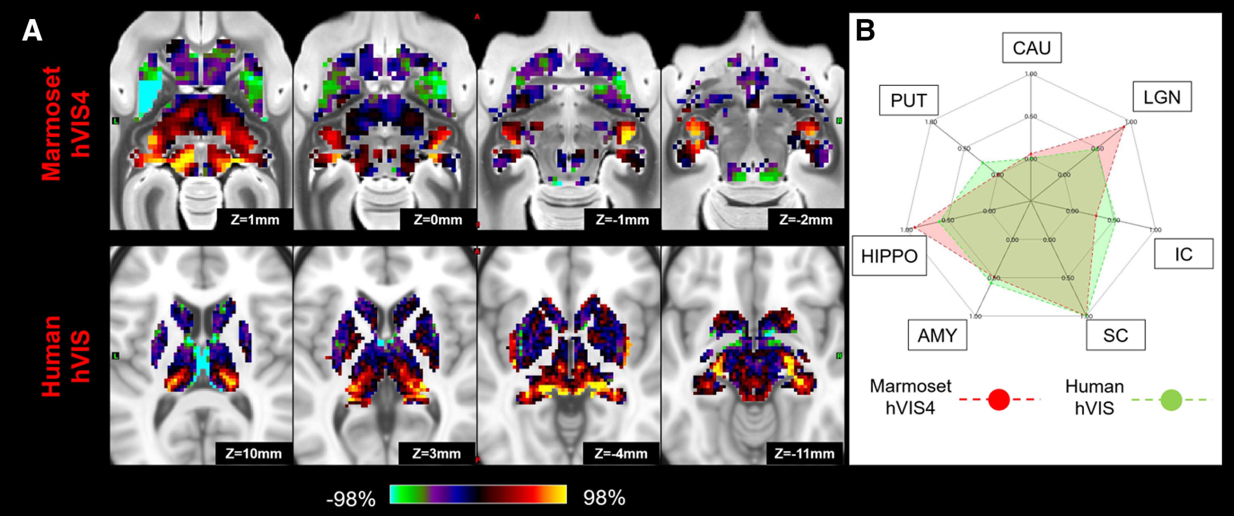
Matching human secondary visual network (hVIS) to marmoset high-order visual network (hVIS4) in subcortical area. A, z score maps were shown in axial slices focused on the superior colliculus and LGN, which have strong connections in both species. A single-color palette applies to two species but is scaled according to percentile ranges within each species rather than to absolute values. B, A fingerprint shows the matching connectivity patterns between marmosets and humans. Red and green areas represent marmoset and human fingerprints, respectively. CAU, Caudate; PUT, putamen; HIPPO, hippocampus; AMY, amygdala.