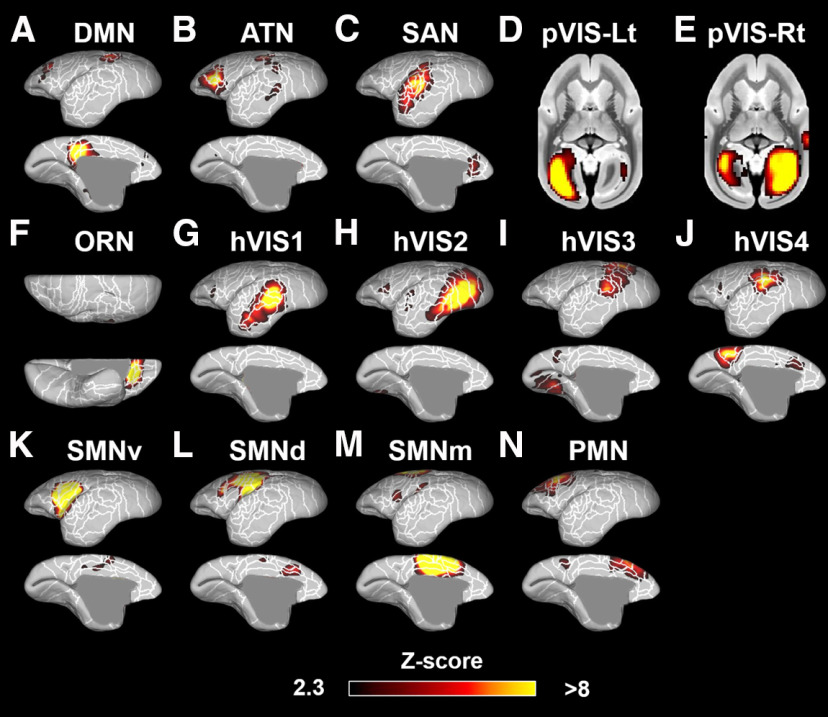
Figure 3.
Fourteen components identified as RSNs in the marmosets. These networks were labeled based on previous studies (Belcher et al., 2013; Hori et al., 2020b) as follows: A, DMN. B, ATN. C, SAN. D, Left primary visual network (pVIS-Lt). E, Right primary VIS (pVIS-Rt). F, ORN. G–J, hVIS1-4. K-M, Somatomotor networks ventral (SMNv), dorsal (SMNd), and medial (SMNm). N, Premotor network (PMN). Color bar represents the z score of these correlation patterns thresholding at 2.3. White lines indicate the cytoarchitectonic borders for reference (Liu et al., 2018).

