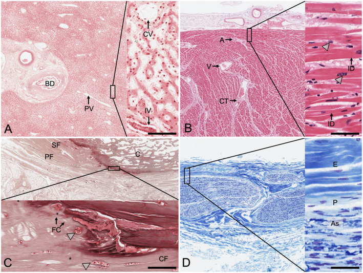
Figure 3.

Histological samples showing the results from phenoxyethanol‐based embalming following a 10‐month duration of the embalmed tissues in the dissection course. The highlighted squares on the left are magnified as inserts on the right side of each image set. A, Liver (reticulin stain). The classical arrangement of hepatic lobules can be seen, with the central veins (CV) and the periportal space. The staining shows well‐preserved tissues, but the fibers appear to mask the fixative. BD, bile duct; IV, interlobular vein; PV, branch of portal vein; scale bar 40 μm. B, Cardiac muscle (H&E stain). This tissue sample from the left ventricle shows intact cardiac myocytes and at higher magnifications intercalated disks (ID). The arrowheads indicate the nuclei of the cardiac myocytes. A, arteriole; CT, connective tissue; V, vein; scale bar 25 μm. C, Bone, calcaneus (silver stain). The calcaneal insertion of the plantar fascia (PF) with the adjacent superficial foot muscle layer (SF) is presented in sagittal orientation. The arrowheads point at the chondrocytes forming the fibrocartilage (FC) at the transitional zone of the collagen fiber (CF) insertions. Only remnants of osteocytes can be seen within the osteon structure of the calcaneus (C). No canaliculi become visible following the staining; scale bar 25 μm. D, Peripheral nerve (Luxol fast blue/cresyl violet stain). The myelin is stained in the various bundles of axons in this sample of the lateral cutaneous nerve of the thigh. Axons (As) and nuclei of Schwann cells are visible. E, epineurium, P, perineurium; scale bar 50 μm.
