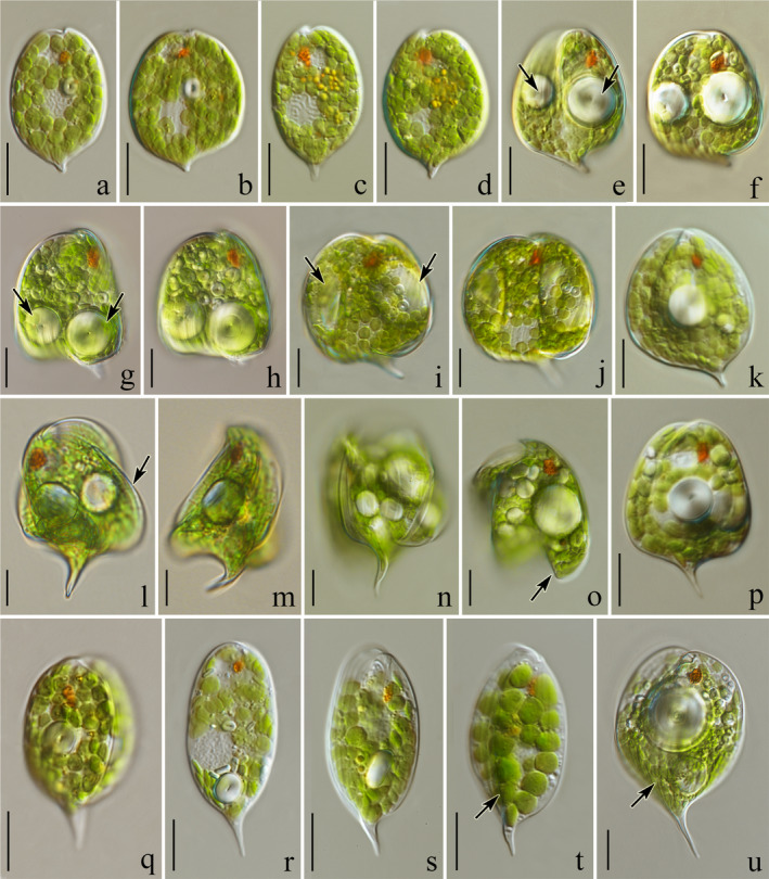
Fig. 3.

Light microscope photographs showing an overview of living cells of the studied Phacus strains (=isolates): (a, b) oval, leaf‐like flat cells of Phacus acuminatus (strain UTEX 1317) terminated with a short, sharp, wedge‐shaped tail; (c, d) cells of P. minutus (strain ACOI 1755) ending with a tail that is bent toward the dorsal side; (e, f) P. curvicauda (isolate UW2262INo), cells with centrally located, large, spherical paramylon grains (arrows); (g, h) trapezoid‐shaped cells of P. anomalus (isolate UW1984Cie) with two large, spherical paramylon grains located in the widest and thickest bottom part (arrows); (i, j) two large, parietal, ring‐like paramylon grains visible in the cell of P. alatus (strain CCAC 2423 B); (k) P. orbicularis (isolate UW2362 Choc); (l–o) spherical cells of P. anacoelus (isolate UW2219IK1) with visible crests (arrows) and large, spherical paramylon grains located in the center; (p) trapezoid‐shaped cell of P. pleuronectes (isolate UW2211Jel); (q) rotund (triangular when cross‐sectioned) cell of P. ankylonoton (strain CCAC 0043) terminated with a straight tail; (r) flat, elongated oval‐like cells of P. applanatus (strain CCAC 2604 B) ending with a straight tail; (s, t) cells of. P. caudatus (strain CCAC 0034) with a dorsal crest which elongates into a sharp hyaline tail slightly bent toward the ventral side; (u) wide‐oval cell of P. manginii (isolate UW2219IK1) with a low crest which elongates into a sharp hyaline tail bent slightly sideways. Scale bars 10 µm.
