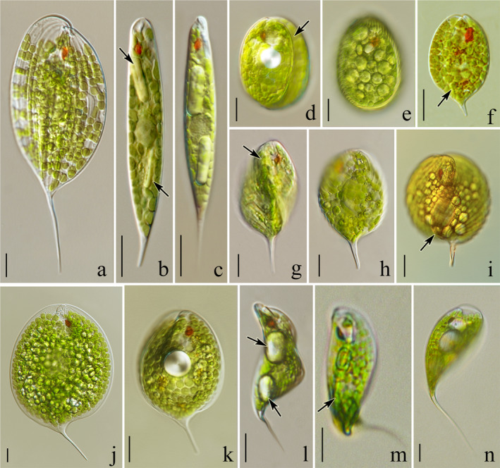
Fig. 4.

Light microscope photographs showing an overview of living cells of the studied Phacus strains (=isolates): (a) elongated oval‐like cell of Phacus elegans (isolate UW2064Ur19) elongated with a long, thorn‐like, straight tail; (b, c) spindle‐shaped cells of P. limnophilus with two large, rod‐like paramylon grains visible, (b) strain ACOI 1026, (c) isolate UW1988Reg; (d) wide‐oval cell of P. stokesii (isolate UW2399Ur19) rounded at both ends; (e) spherical cells of P. salinus (strain SAG 1244‐3); (f) P. tenuis (strain ACOI 1757), oval cell with a low crest elongated into a tail bent slightly toward the ventral side; (g–i) almost spherical, slightly twisted cells of P. arnoldii with three ridges (g, h) strain CCAC 2432B, (i) isolate UW2313Pil; (j) large, flat, almost round cell of P. gigas (isolate UW1669Ora) terminated with a sharp tail bent sideways; (k) spoon‐shaped (convex) cell of P. hamatus (strain CCAC 2605 B [=ASW 08032]) ending with sharp hyaline tail bent visibly toward the ventral side; (l–m) cylindrical, flat, slightly U‐bent and spirally twisted cells of P. raciborskii (isolate UW2326Gra2); (n) cell of P. lismorensis (isolate UW1930Dol) bent like a bow, terminated with a long, sharp, pointy tail. Scale bars 10 µm.
