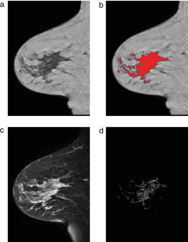
FIGURE 2.

Example of the image processing from cohort 2 in the contralateral breast of a 42‐year‐old patient with an ER‐positive/HER2‐negative invasive ductal carcinoma in the right breast. (a) Sagittal T1‐weighted image. (b) Same image with parenchymal tissue overlaid in red. (c) Fat‐suppressed first postcontrast image. (d) Enhancement in the parenchymal tissue segmentation.
