
Figure 3.
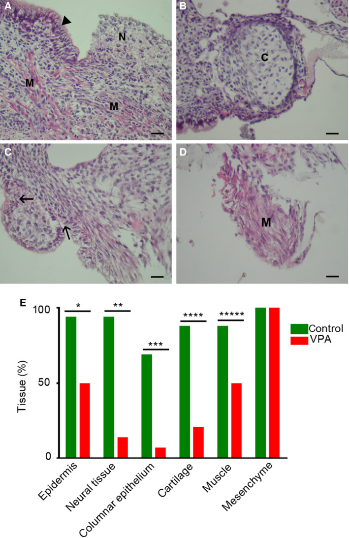
Differentiation in embryo‐derived teratomas cultivated for 14 days in vitro. (A, B) Control trilaminar embryo‐derived teratomas cultivated in Eagle's MEM and 50% rat serum. Pseudostratified columnar epithelium (arrowhead); neural tissue (N); muscle (M), cartilage (C), scale bar = 25 μm, hematoxylin–eosin. (C, D) Embryo‐derived teratomas cultivated in Eagle's MEM and 50% rat serum with 2 mm VPA. Cylindrical epithelium (arrows); muscle (M), scale bar = 25 μm, hematoxylin–eosin. E Incidence of tissues (%) in the group of control embryo‐derived teratomas (n = 16) and in the group of VPA‐treated embryo‐derived teratomas (n = 14). Fisher's exact test, with two‐tailed P calculation. *P = 0.0121, **P = 0.0001, ***P = 0.0008, ****P = 0.0006, *****P = 0.0457.