
Figure 4.
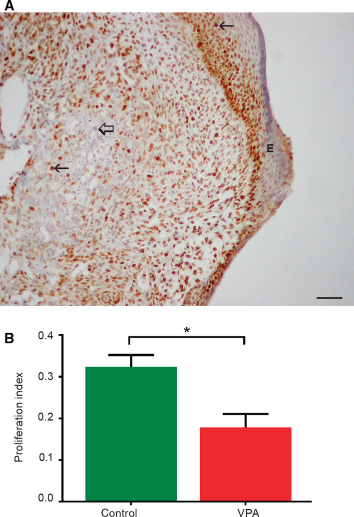
Expression of the PCNA in embryo‐derived teratomas cultivated for 14 days in vitro. (A) PCNA‐positive cells (arrows) in control teratomas; negative internal control (thick arrow), epidermis (E), scale bar = 50 μm. IHC, DAB, counterstained by hematoxylin. (B) Proliferation index (NPCNA/800 cells; n = 14 control embryo‐derived teratomas, n = 11 VPA‐treated embryo‐derived teratomas). Mean ± SEM. *P = 0.0055 (Student's t‐test, two‐tailed).