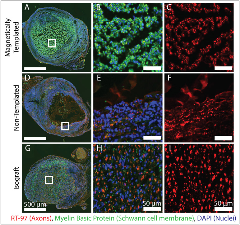
Figure 7. Axonal Elongation and Schwann Cell Migration into the Proximal End of Experimental Implants After 4-Weeks of Implantation.
A-I) Fluorescence stained images of magnetically templated, non-templated, and isograft implants taken at the proximal implant location 2 mm into the device after 4-weeks in vivo. Left panel shows complete cross sections, with magnified views shown to the right as indicated by white boxes. Left panel shows RT97 staining for axons, and all other images are stained for RT97 for axons (red), Myelin basic protein (green), and DAPI (blue). (n = 2 / group). At the proximal graft location, isografts and magnetically templated hydrogels demonstrated axonal growth and Schwann cell infiltration in the luminal area, compared to minimal axonal growth localized to the hydrogel-SIS interface in non-templated samples.

