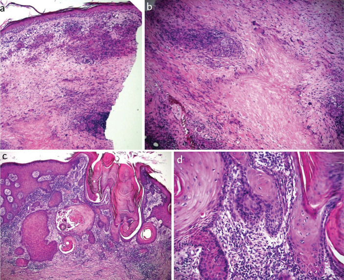
Fig 2.
Haematoxylin and eosin stains showing an abnormal squamous proliferation composed of small islands with irregular edges infiltrating a chronically inflamed and fibrotic dermis in keeping with invasive well differentiated squamous cell carcinoma. a) At ×10 magnification. b) At ×20 magnification. c) At ×10 magnification. d) At ×40 magnification.

