Fig. 4.
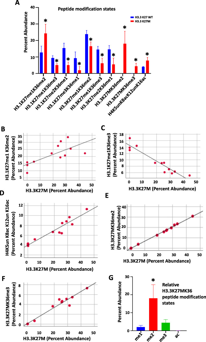
Distinct peptide modification states detected in DIPG tumor tissue. a Distinct combinations of modification states are observed on ten peptides in H3.3K27M mutant tumor tissue (n = 9) relative to H3.3K27 wild-type (WT) tissue (n = 3).* p < 0.05 compared to H3.3K27 WT group, Independent-sample t test, two-tailed. b-f Scatter plots showing the correlation between relative abundance of H3.1K27me1K36me2/me3, H4K5un K8acK12unK16ac, and H3.3K27MK36me2/3 peptides with the H3.3K27M peptide in DIPG tumor tissue (Pearson Correlation = 0.72, −0.79, 0.95, 0.99, and 0.97, respectively, all p < 0.01. Pearson Correlation, two-tailed, n = 12). g Relative abundance of H3.3K27MK36 peptide modification states in DIPG tumor tissue reveals K36me2 as the most abundant co-occurring peptide with H3.3K27M mutation peptide. * p < 0.05 compared to all the other groups (Tukey HSD, Post Hoc Tests, one way ANOVA, n = 9)

