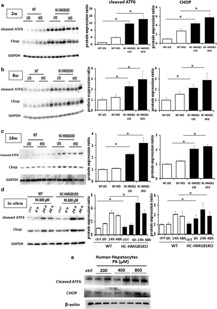
Fig. 4.
ER stress in WT and HC-HMGB1−/− (KO) liver up to 16 weeks after LFD or HFD feeding, and in hepatocytes after PA treatment. Western blot analyses of levels of cleaved ATF6 and CHOP (ER stress markers) in WT and HC-HMGB1−/− (KO) liver at a 2 weeks, b 8 weeks and c 16 weeks after LFD or HFD feeding. Lysates from one mouse liver per lane. d Western blot of WT and HC-HMGB1−/− (KO) hepatocyte whole cell lysates in control (ctrl—no treatment), or time points up to 36 h after PA treatment. Gray analyses was performed for quantization of protein expression. Three independent experiments were performed. All data are means ± SEM. *P < 0.05. e Western blot of human hepatocyte whole cell lysates in control (ctrl—no treatment), or 24 h after PA treatment. GAPDH and β-actin were used as a loading control. Images representative of at least three repeated experiments

