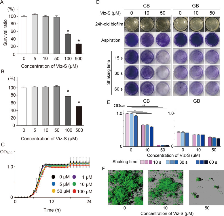
Fig. 1.
Cytotoxicity of Viz-S in (a) human gingival epithelial cells (HGECs) and (b) human gingival fibroblasts (HGFs). Cells were incubated in MEM or DMEM containing Viz-S for 3 h at 37 °C. The cellular viability was assessed using an MTT assay (n = 6). *p < 0.01, as compared with the control group. c Growth curves of bacteria derived from human saliva in the presence or absence of Viz-S (n = 4). There were no significant growth differences among the concentrations of Viz-S (p > 0.05). d Residual biovolumes following shaking motion for 15, 30 and 60 s (n = 5). The remaining structure was stained with 0.1% crystal violet. A 24h-old biofilm: Photographs of the biofilm structure after a 24-h incubation. Aspiration: The structure after the supernatant was removed without a washing procedure. e Total biomass determined by measuring absorbance at 570 nm (n = 5). **p < 0.01, *p < 0.05, compared with the control group. f Three-dimensional reconstruction images of residual CB stained with a fluorescent bacterial viability kit following shaking motion for 15 s (n = 3). Live bacteria appear fluorescent green (SYTO9) and dead bacteria appear fluorescent red (PI). Scale bar = 30 μm

