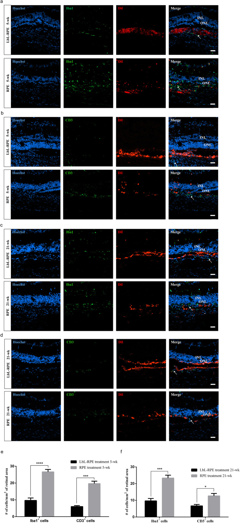
Fig. 7.

Immunogenicity of RPE cells or LbL-RPE cells at 5 and 21 weeks after transplantation. a–d Photomicrographs showed the labeling of RCS rats’ retinal sections at 5 and 21 weeks after transplantation. Anti-Iba1/CD3 antibody (green); many Iba1+ cells (arrow) invaded the INL/ONL after RPE transplants, but were poorly labeled after LbL-RPE transplants (RPE and LbL-RPE cells were pre-labeled with Dil (red)). There were numerous CD3+ cells (arrow) which infiltrated in the RPE retinas. CD3+ cells were very sparse in LbL-RPE transplants. Scale bars 20 μm. e, f The number of positive Iba1+ and CD3+ cells around the transplanted retinas at 5 and 21 weeks after transplantation (mean ± SEM; ****p < 0.0001; ***p < 0.001; *p < 0.05; n = 8)
