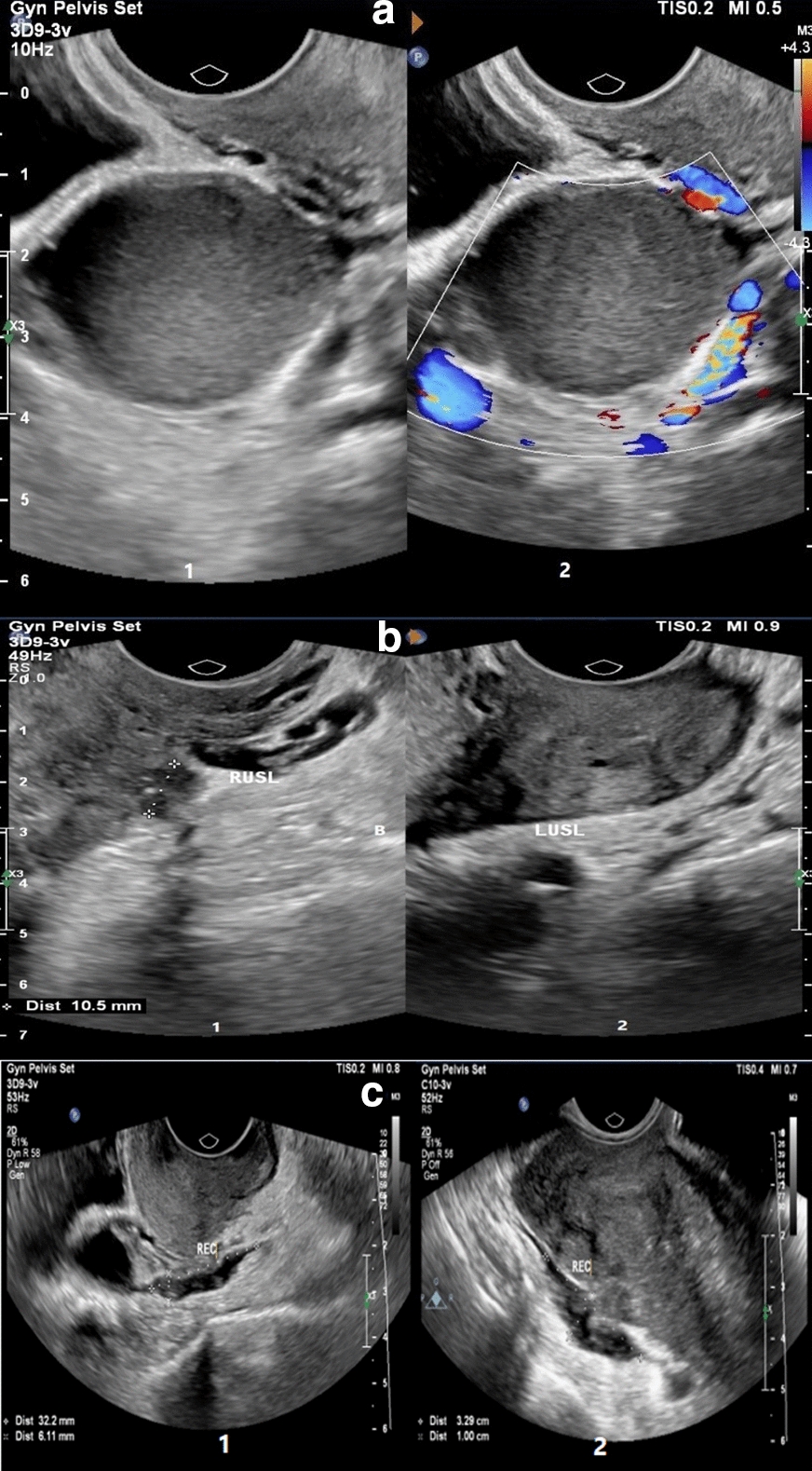
Fig. 1.

a Typical ovarian endometrioma in a woman with long-standing chronic pelvic pain and dysmenorrhea (1, 2) gray-scale (1) and color Doppler (1) TVS images of right ovary demonstrate a unilocular cyst containing homogeneous low-level echoes and no internal vascularity at color Doppler US (classic appearance of an ovarian endometrioma). b USL DIE in a woman with severe pelvic pain and dyspareunia for 10 years with a history of stage IV endometriosis who was confirmed to have extensive endometriosis at laparoscopy. (1) sagittal gray-scale TVS image shows irregular thickening of the right USL associated 10 mm endometriosis nodule in proximal. (2) Also, a moderate thickening of left USL has been shown. c Bowel DIE in two women. (1) Sagittal gray-scale TVS image in a woman with severe dysmenorrhea shows a hypoechoic nodule involving the serosal layer in the lower rectum. (2) Transverse gray-scale TVS images in a woman with chronic pelvic pain and cramping, show a hypoechoic nodule in the rectosigmoid junction with severe adhesion to the posterior of the uterus fundus
