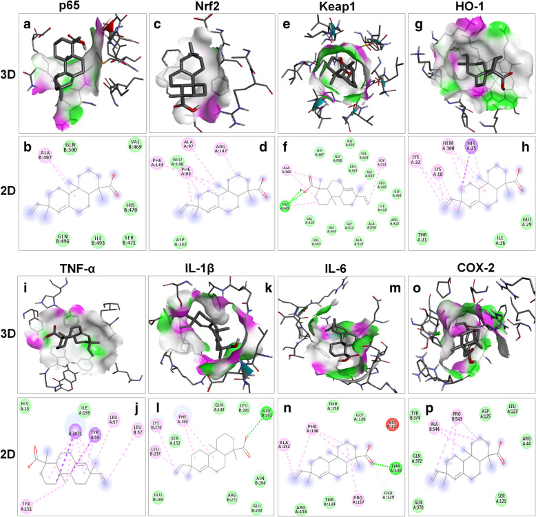
Fig. 7.
The binding interaction of the continentalic acid with various protein target such as p65 (PDB-ID: 1vkx), Nrf2 (PDB-ID: 2flu), Keap1 (PDB-ID: 4iqk), HO-1 (PDB-ID: 1ubb), TNF-α (PDB-ID: 2az5), IL-1β (PDB-ID: 1itb), IL-6 (PDB-ID: 1p9m) and COX-2 (PDB-ID: 5ikq) with their subsequent 3D and 2D structure. The docking analysis was done to calculate the binding energy, vander wall forces and type of binding. The interaction of the continentalic acid with the p65 (a, b), Nrf2 (c, d), Keap1 (e, f), HO-1 (g, h), TNF-α (i, j), IL-1β (k, l), IL-6 (m, n) and COX-2 (o, p) was shown in both 2D and 3D view. The 3D view show the binding pocket within the ligand orient, while the 2D images show the interactive amino acids interacting with the ligand and type of interaction

