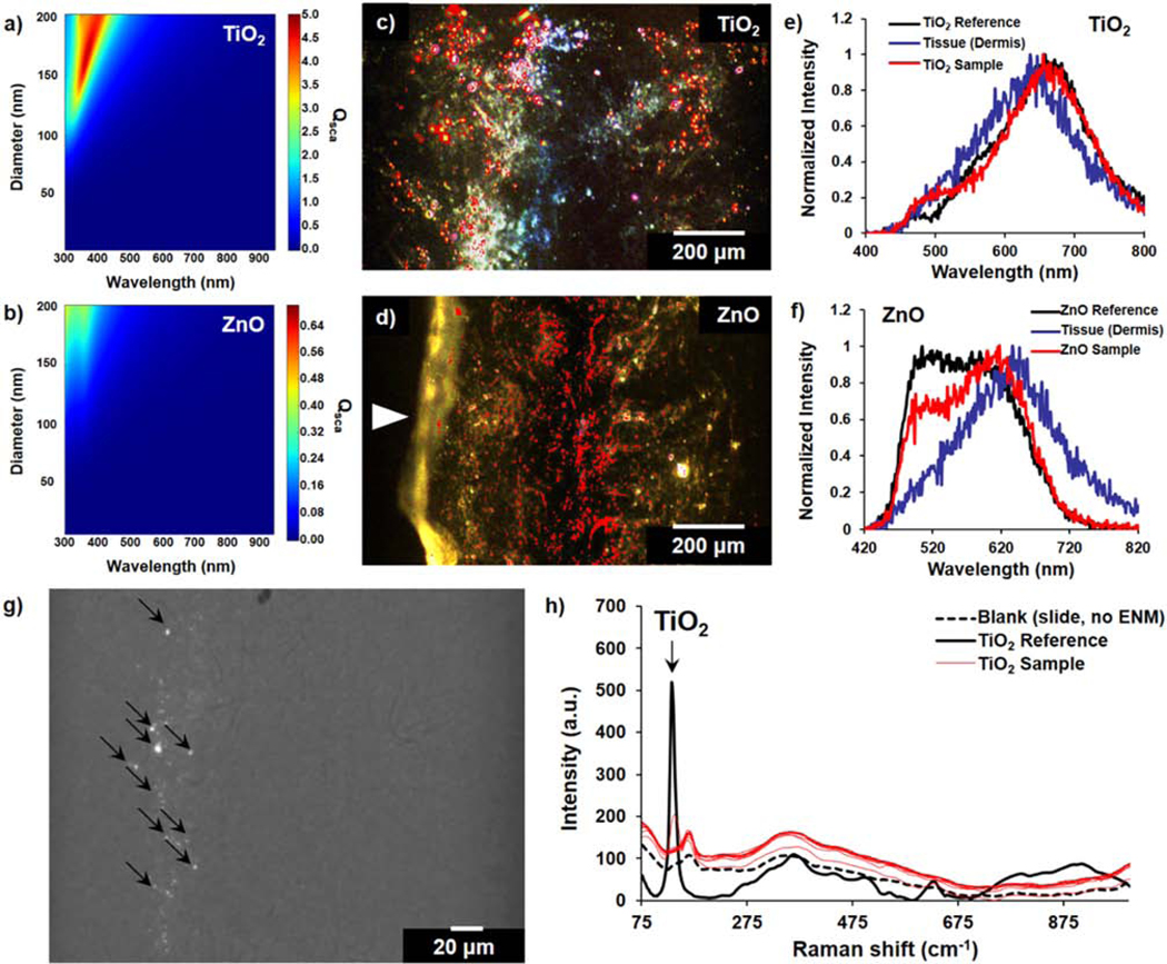
Figure 5. Label-free mapping of TiO2 and ZnO within skin tissue sections using micro-spectroscopy techniques.

(a, b) Calculated scattering efficiency (Qsca) of TiO2 and ZnO at different diameters and multiple wavelengths, based on Lorenz-Mie theory model of particle scattering. (c, d) Enhanced darkfield images of ENM-exposed skin tissue sections overlaid with (c) TiO2 and (d) ZnO distribution map; annotated as red highlighted regions. White arrow in (d) indicates the epidermal region. (e, f) Representative scattering spectra taken from hyperspectral scans of tissue samples compared to a reference slide with TiO2 or ZnO aqueous solutions and background dermal tissue signal. (g) Darkfield-visible TiO2 ENMs in cleared skin sections; merged with brightfield image. (h) Raman scans of points in a representative cleared skin section with darkfield-visible scattering signal; compared to a reference TiO2 and blank glass slide. Black arrows in (g) represent the spots corresponding to the reported Raman spectra in (h). For these 24 h exposure experiments, only the dermal side of the biopsies was exposed to ENM suspensions.
