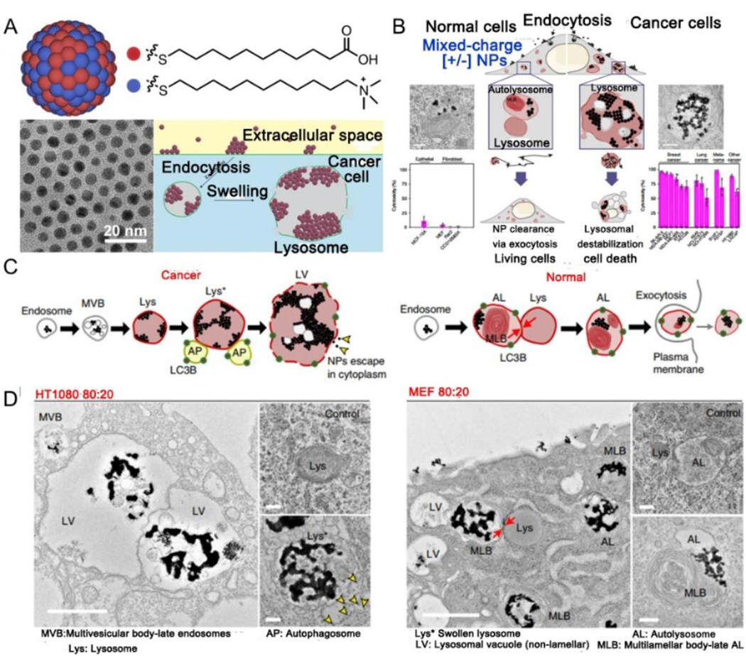
Fig. 19.

(A) Schematic structure and pH-responsive cascaded aggregation of mixed-charge NPs ([+/−] NP). (B) Summary of how crystallization of [+/−] NP in lysosomes results in selective destroying of cancer cells. (C) Proposed aggregation mechanism of cancer (left) versus normal (right) cells. (D) TEM images of HT1080 (left) or MEF (right) cells at 24 h post-treated with 4:1 NPs. (A-D) Reproduced with permission.[281–282] Copyright 2020, Springer Nature Limited.
