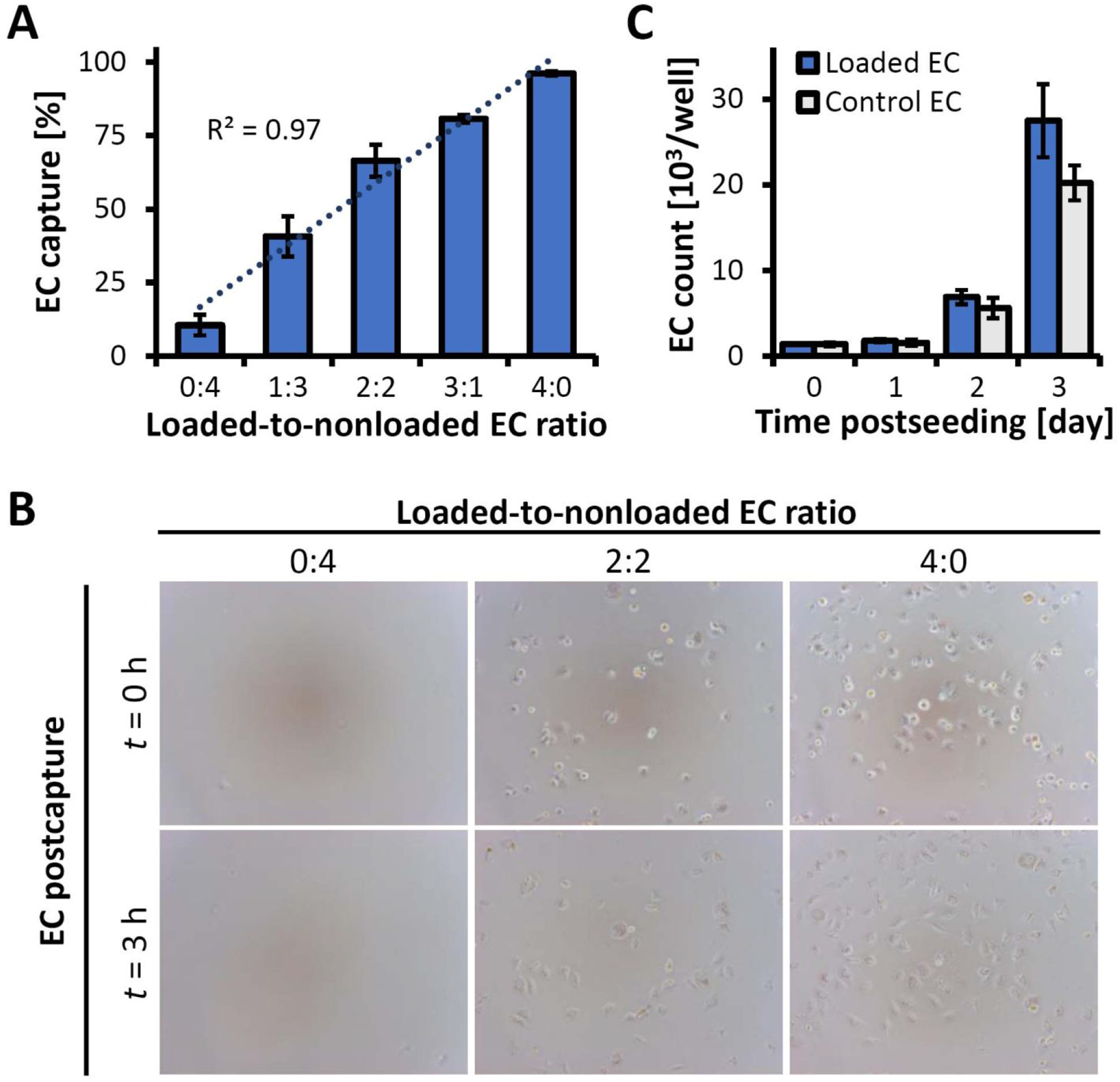
Figure 2.

Characterization of magnetic responsiveness, substrate binding stability and EC growth using the inverted-plate assay. (A) EC capture from binary suspensions of MNP-loaded and non-loaded EC showing selective and quantitative separation of magnetically responsive cells at all tested ratios. (B) Representative micrographs of EC captured from binary mixtures immediately following capture (t = 0 hr) and 3 hr post-capture, showing magnetically driven substrate attachment and stable anchorage (magnification ×100). (C) The number of magnetically captured, functionalized EC (“Loaded EC”) 0–3 days post-seeding, shown in comparison to non-functionalized control cells seeded at the same initial density (“Control EC”). Statistical analysis shows no significant difference in the proliferation kinetics (p=0.4).
