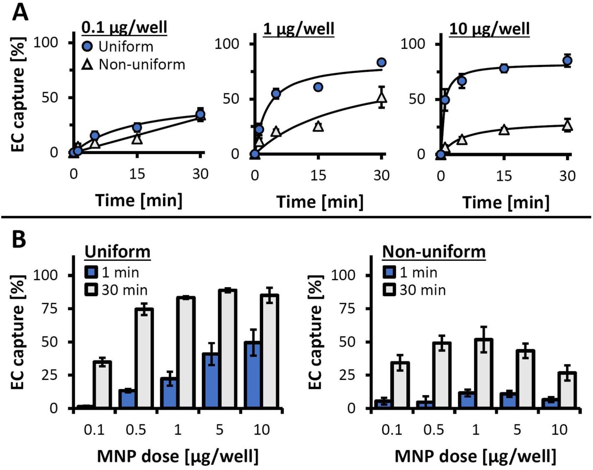
Figure 4.

Magnetic EC capture/anchorage measured longitudinally using the inverted-plate assay as a function of loading protocol and MNP dose. (A) The time courses of EC capture are shown for EC loaded using 0.1, 1, and 10 μg MNP/well with the optimized (uniform) and the simulated, non-uniform loading procedures. EC captured by exposure to the high-gradient magnetic field over different time periods (1, 5, 15, and 30 min) were quantified using the Alamar Blue assay. Data are presented as a fraction of the total cell number (n = 4). (B) Detailed analysis of EC capture/anchorage after 1 and 30 min of magnetic exposure for EC loaded with 0.1–10 μg MNP/well using the optimized (uniform) and the simulated, non-uniform loading procedures.
