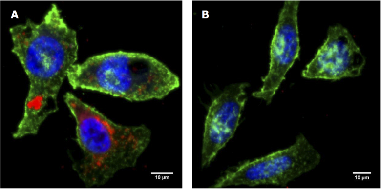
Fig. 10.
Maximum intensity projection of fluorescence microscopy images of HeLa cells incubated with folate conjugated fluorescent magnetic nanoparticles(red) without(A) and with(B) blocking in excess folate. Counterstained with Alexa Fluor 488, WGA membrane stain(green) and Hoechst 33342 nuclear stain(blue).

