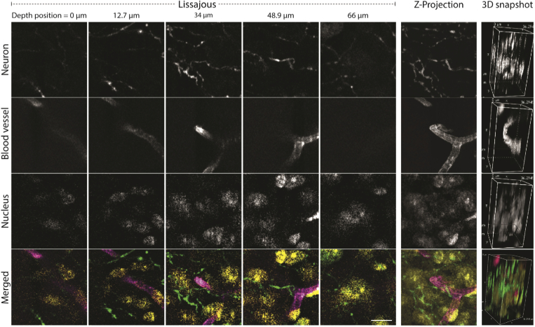
Fig. 3.
Synchronous 3-color volumetric brain imaging. Rows correspond to different color channels, each highlighting a different part of the brain. The bottom row consists of merged images containing the three channels (green for neuron, magenta for blood vessels, and yellow for nuclei). Columns indicate the z-position of the images. The last two right columns correspond to a z-projection and a 3D rendering. The overall axial scan range was 66 µm, and images were reconstructed with 30 scans (2 seconds). A video of the 3D rendered images is available (Visualization 1 (4.5MB, avi) ). Fluorescent staining: Alexa Fluor 488 for blood vessels excited at 488 nm, SYTOX Orange for nuclei at 561 nm, and Alexa Fluor 633 for neurons at 640 nm. Scale bar 10 µm, Voxel size 0.062 × 0.062 × 2.4 µm3, fTAG = 457141 Hz, Lissajous period 2π.

