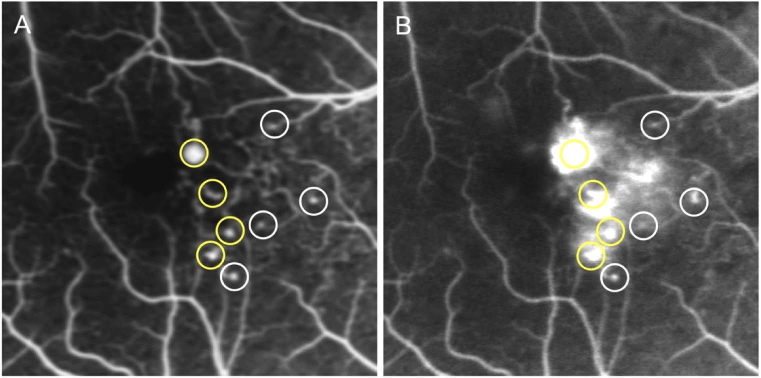
Fig. 1.
Detection of microaneurysms (MAs) and the evaluation of the associated-dye leakage using fluorescein angiography (FA). FA images obtained at the early (A) and late (B) phases. The MAs are shown as hyperfluorescent dots in the early phase. Fluorescein leakage is defined as any increased intensity over the choroidal background, within the retina, but outside the retinal vasculature. Yellow and white circles indicate MAs with and without significant leakage, respectively.

