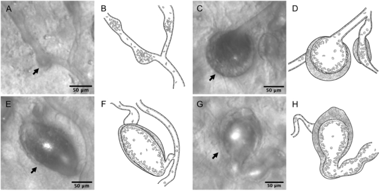
Fig. 3.
Morphological classifications of microaneurysms (MAs) associated with retinal vein occlusion. (A-B) Focal bulge type (arrow). (C-H) Non-focal bulge type, which are larger than non-focal bulge MAs. (C-D) Saccular type (arrow). (E-F) Fusiform type (arrow). (G-H) Mixed type (arrow). (B,D,F,H) Illustrations of each Figure of first and third column, respectively.

