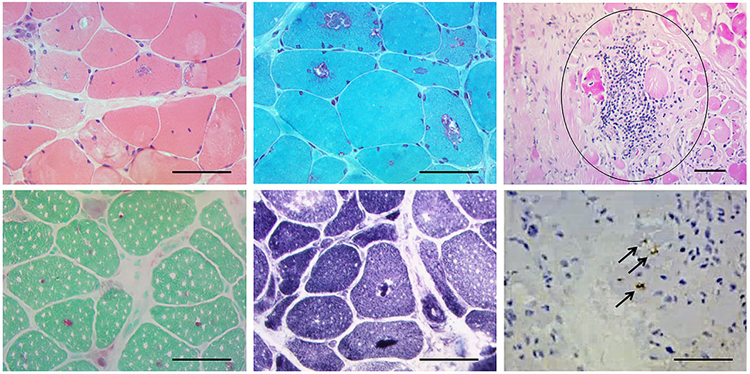
FIGURE 1.

Muscle biopsy showing central and sub-sarcolemmal vacuoles in hemotoxylin and eosin (H&E) (top panel left), which demonstrates red “rimming” with modified Gomori trichrome (top center panel). An example of inflammation is seen in the top right panel (circled). Bottom panel: Acid phosphatase (left) and NADH (center) showing positive material within the vacuoles and vacuoles are stained positive for ubiquitin (black arrows) (right). Scale bar = 50 μm. Additionally, the connective tissue was mildly increased. Atrophic fibers were round and pyknotic nuclear clumps were not seen, and the biopsy showed a moderate number of fibers with internalized nuclei. Regenerating fibers were not seen. The following stains were normal: cytochrome oxidase (COX), myosin ATPase (normal distribution of fiber types), Oil red O, periodic acid–Schiff (PAS), phosphorylase, Congo red. Neither muscle fiber-type grouping, nor type specific atrophy was seen.
