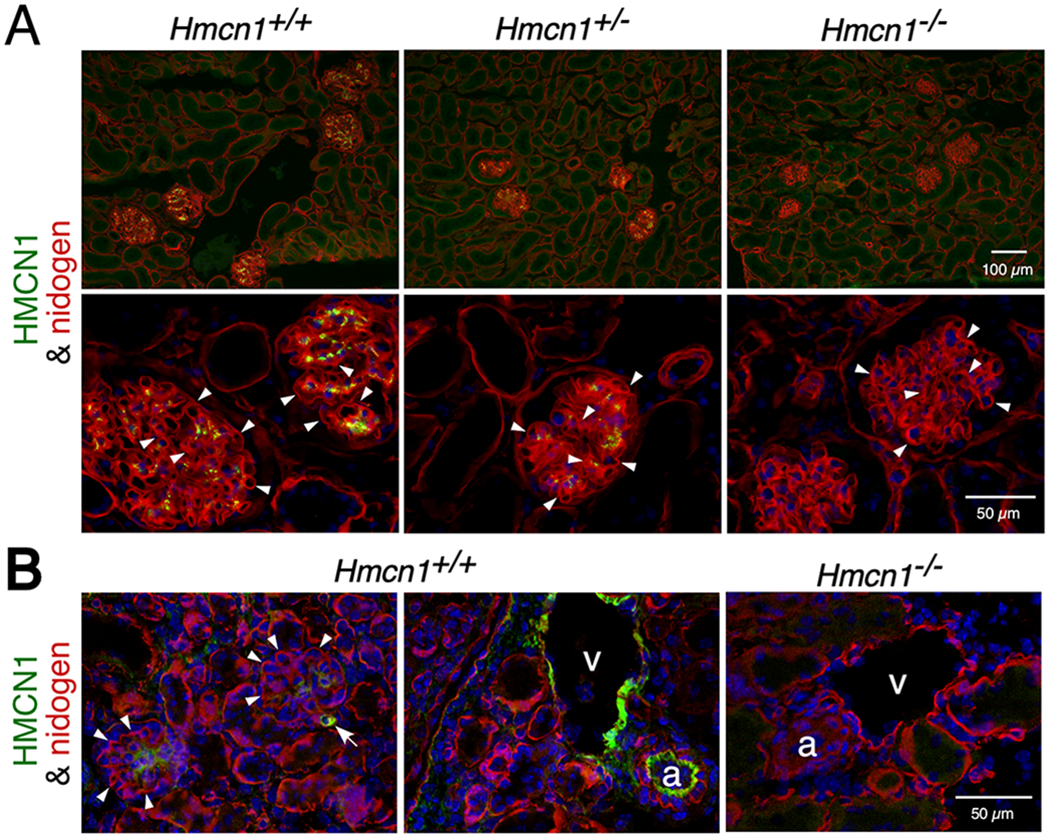
Figure 2. HMCN1 deposition in the kidney.

Kidney sections from mice at 10 months (A) and 2.5 days (B) of age with the genotypes indicated were subjected to immunofluorescence assays. (A) High magnification images are shown in the lower panels. WT and Hmcn1+/− adult kidneys showed deposition of HMCN1 (green) in the mesangial matrix of glomeruli. There was no HMCN1 detected in the nidogen-positive (red) GBM (arrowheads). There were no HMCN1 signals in Hmcn1−/− kidneys, demonstrating the specificity of the antibody. (B) Deposition of HMCN1 was detected in WT pups in the mesangial matrix, small blood vessels (arrow), artery (a), and vein (v) but undetectable in the GBM (arrowheads). Hmcn1−/− kidneys did not show signals.
