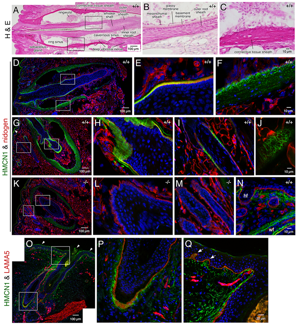
Figure 3. HMCN1 tracks in whisker and hair follicles.

Sections of mouse whisker pads at 8 months (A-M) and 2.5 days (N) of age (Hmcn1 genotypes indicated) and a human skin biopsy (O-P) were subjected to hematoxylin & eosin staining or confocal immunofluorescence assays. The boxed regions in A, D, G, K, O are magnified in the two panels following each of them. HMCN1 was detected in WT adult mice in the BM of the outer root sheath (D, E) and in the connective tissue sheath (F) of whisker follicles, with distinct tracks observed along the BM in sections cut obliquely (G, H). HMCN1 tracks were also detected in hair follicles, along the BM (I) or in a reticular pattern when viewed on the surface of hair follicles (J). There were no specific signals in Hmcn1−/− mice (K, L, M). In addition to hair follicles (hf) and whisker follicles (wf), HMCN1 signals were detected in the dermis of WT whisker pads of pups (N). (O-Q) HMCN1 tracks were found along the BM of human hair follicles (P) and in the dermis and its papillary structures (arrows in Q). Non-specific signals in G and O are marked (arrowheads).
