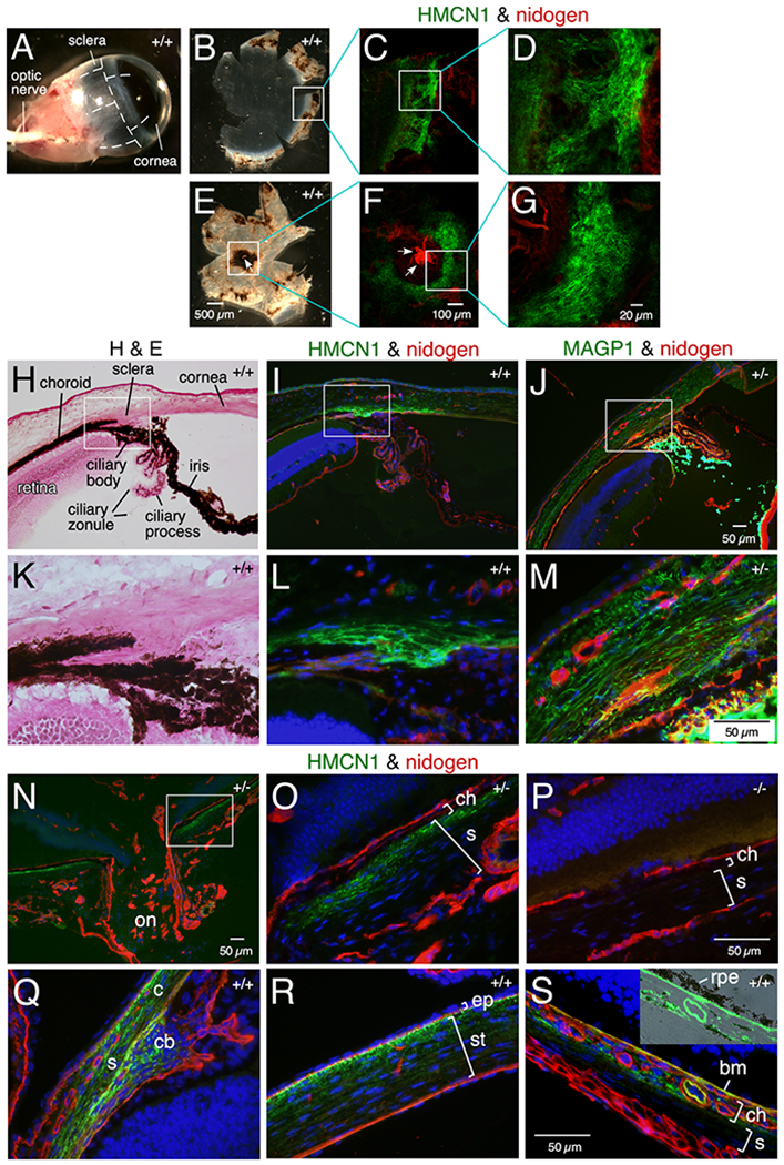
Figure 4. HMCN1 tracks in mouse eyes.

(A-G) WT eyes (11 months) were dissected along the dashed lines (A) to release anterior (B) and posterior (E) eye cups and subjected to immunofluorescence. HMCN1 tracks were detected in scleral regions bordering the cornea (C, D) and surrounding the optic nerve head (ONH) (F, G; arrows point to the ONH). (H-S) Sagittal sections of eyes at age 4 months (H-P) and 2.5 days (Q-S) with Hmcn1 genotypes indicated were subjected to H&E staining or immunofluorescence. The boxed regions in H-J and N are magnified in K-M and O, respectively. Bright HMCN1 tracks were detected in inner scleral layers anterior to the retina (I, L) with weaker signals in the cornea (I), compared to the broader expression domain of microfibril-associated glycoprotein 1 (MAGP1) in sclera and cornea (J, M). Bright MAGP1 staining marks ciliary zonules. The HMCN1 tracks detected around the ONH (on in N) were confined to the inner scleral layer (s) abutting the choroid (ch in O). There were no HMCN1 signals in Hmcn1−/− mice (P). In WT pup eyes, HMCN1 tracks were detected in scleral regions (s) bordering the cornea (c), in the ciliary body (cb in Q), in corneal stroma (st) with stronger signals toward the corneal epithelium (ep in R), in the choroid (ch), and in Bruch’s membrane (bm), but tracks were undetectable in remaining scleral regions (s in S). Inset in S is an overlay of HMCN1 signals with the bright-field image, showing the HMCN1-stained Bruch’s membrane abutting the retinal pigment epithelium (rpe).
