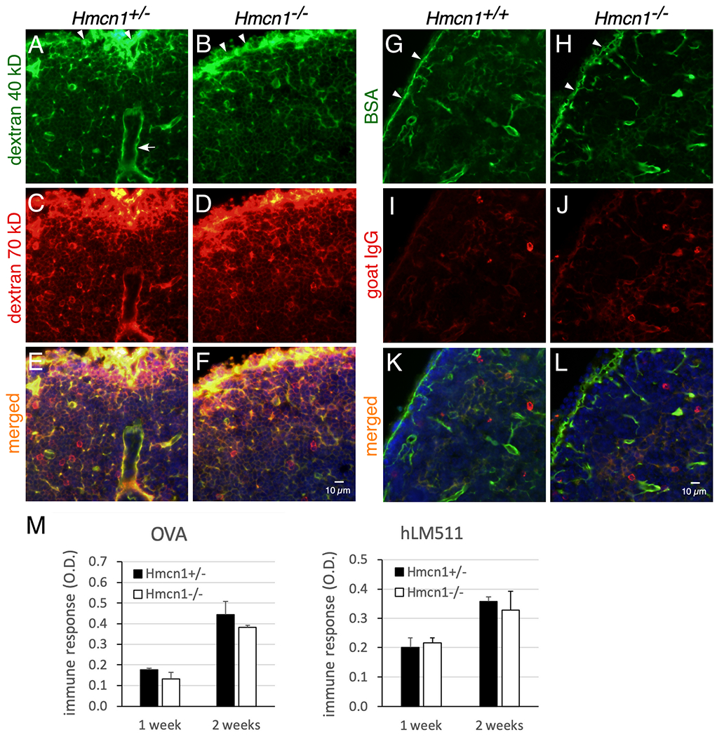
Figure 7. Analyses of effects of HMCN1 deficiency by tracer distribution and immune responses.

(A-L) Mice of the indicated genotypes at 7 to 9 months of age were injected subcutaneously into footpads with a mixture of 40-kD dextran-fluorescein and 70-kD dextran-TMR (A-F) or a mixture of BSA-FITC (68 kDa) and goat anti-rabbit-IgG-Cy3 (150 kDa) (G-L). Popliteal (A-F) and brachial (G-L) lymph nodes were collected at 15 and 9 min after injection, respectively, fixed, and then sectioned. (A-F) Injected 40- and 70-kDa dextrans were distributed in the subcapsular sinuses (arrowheads) and in conduits of B-cell follicles in both control and mutant mice. (G-L) Injected BSA was also distributed to subcapsular sinuses and to conduits of B-cell follicles. In contrast, the distribution of goat IgG in the same lymph node was weak (I-L). Similar results were obtained for control and mutant mice. Arrow in A, high endothelial venule. (M) Mice at 9 to 10 months of age were immunized by intraperitoneal injections with ovalbumin (OVA) or human laminin-511 (hLM511) daily for 3 days. By ELISA control and Hmcn1−/− mice showed similar induction of antibodies to OVA or hLM511 at 1 and 2 weeks of immunization.
