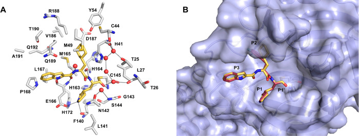
Figure 7.
Co-crystallographic pose of compound 5 (yellow-orange sticks, PDB 6LZE) covalently bound to the active site of SARS-CoV-2 3CLpro. (A) The key residues forming the binding pocket are displayed as white sticks; water molecules are shown as red spheres. H-bonds are depicted as dashed black lines. (B) Overlay of 5 and 6 (raspberry sticks, PDB 6M0K) co-crystallographic poses.

