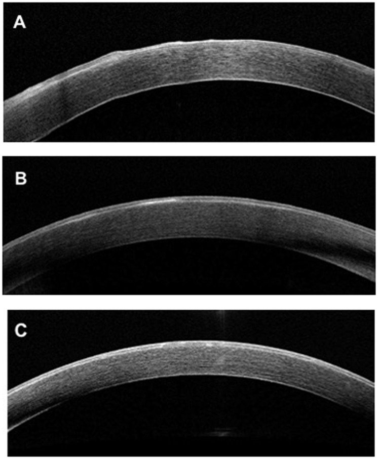
Fig 2. Spectral-domain optical coherence tomography B-scan of cornea with EBMD.

A: Presence of an irregular and thickened epithelial basement membrane with duplication associated with undulation and elevation of the corneal epithelial layer. B: Presence of a thickened and hyperreflective basement membrane. C: Presence of hyper-reflective dots in the middle of the corneal epithelial layer.
