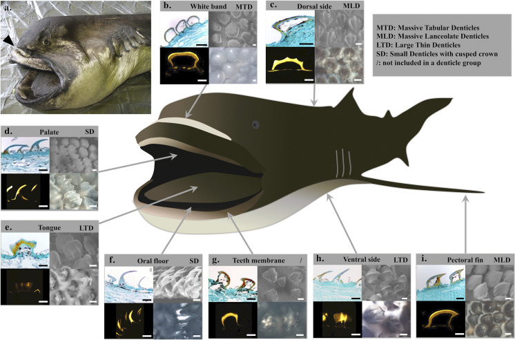
Fig 2. Histological analyses of the megamouth, Megachasma pelagios, skin with an emphasis on denticles.
(a) Specimen of M. pelagios (OCA-P 20110301) examined during this work. Arrowhead indicates the white band visible when the jaw is protruded. Histological approaches of the (b) white band, (c) dorsal skin, (d) palate, (e) tongue, (f) oral floor, (g) teeth membrane, (h) ventral skin, (i) pectoral skin. Results are represented for each skin zone: paraffin section (upper left), SEM micrography (upper right), microradiography (lower left), and transmitted light microscope upper view (lower right). Scale bars: 100 μm.

