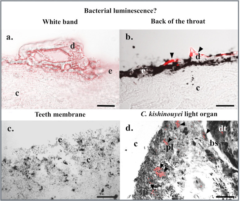
Fig 3. Fluorescence in situ hybridization: Bacterial luminescence?
Fluorescence in situ hybridization on (a) white band, (b) back of the throat, and (c) teeth membrane skin sections of M. pelagios as well as (d) C. kishinouyei light organ sections using EUB RNA probes. Black arrowheads indicate bacteria-labeled areas. bs, basal layer of the digestive tract-related tubules; c, connective tissue; d, dermal denticle; dt, digestive tract tubule; e, epidermis; pl, pigmented layer. Scale bars: 100 μm.

