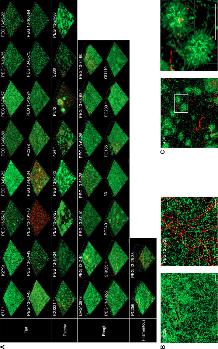
FIG 2.
High-level architectural heterogeneity in 40 different clinical S. maltophilia isolates. Biofilm cells were grown under flow or static conditions for 72 h. After a LIVE/DEAD staining, the biofilm architectures were recorded using CLSM. Red, dead cells; green, living cells. (A) The isolates were grouped as forming flat, rough, patchy, and filamentous biofilms based on their overall architectures. Strain identifiers are indicated on the top left corner for each isolate. Strains used in additional transcriptome data are marked with an asterisk. Images represent an area of 100 μm by 100 μm of the respective biofilm. For each of the 40 isolates, at least 3 areas were analyzed. (B) Multicellular and filamentous forms of the isolates PC256 and PEG 13-25-38 are shown via a top view on the biofilm architecture. Scale bar represents 10 μm. (C) Isolate 454 forms rosette-like multicellular clusters of cells. In the right panel, a 4-fold magnification of the boxed area of the left panel is depicted. Scale bar represents 10 μm.

