
Fig. 3. Heterogeneous micro/nanofibers could affect the expression of integrin β1 and vinculin.
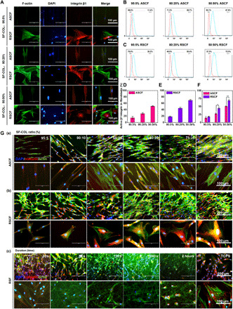
(A) Immunofluorescence staining of integrin β1 in fibroblasts cultured on ASCF and RSDF containing varying COL-I content (5, 20, and 50%). DAPI, 4′,6-diamidino-2-phenylindole. (B and C) Flow cytometry of fibroblasts cultured on ASCF and RSCF containing varying COL-I content (5, 20, and 50%). (D) Integrin β1 activation rate of fibroblasts on ASCF. (E) Integrin β1 activation rate of fibroblasts on RSCF. (F) Comparison on integrin β1 activation rate of fibroblasts on ASCF and RSCF under various COL-I ratios (5, 20, and 50%). Statistically significant differences were indicated by *P < 0.05 and **P < 0.01 when comparing between ASCF and RSCF groups. (G) Immunofluorescence staining of vinculin in cells cultured on different fibers. (a and b) Immunofluorescence staining of vinculin in fibroblasts cultured on (a) ASCF and (b) RSCF containing gradient COL-I ratios, heterogeneous microstructure, and chemical cues. (c) Immunofluorescence staining of vinculin in myofibroblasts cultured on RSF with different fiber densities.