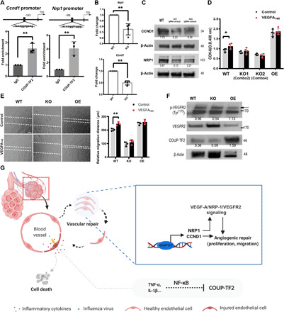
Fig. 6. COUP-TF2 directly targets CCND1 and NRP1 to mediate cell proliferation and migration.

(A) ChIP followed by qPCR was performed to amplify the target region in the Ccnd1 and Nrp1 promoters. (B) qPCR analysis of COUP-TF2 in WT and COUP-TF2 KO iMVECs. (C) Immunoblotting of CCND1 and NRP1 in both WT and KO iMVECs, numbers under Western blot bands represent relative quantifications over actin. n = 2. (D) CCK-8 assay showing cell proliferation of iMVECs treated ±VEGFA165 (20 ng/ml) for 72 hours. n = 4. (E) Cell migration was assessed using a wound scratch assay. Images were obtained at (0 hours) and (24 hours). Representative photos illustrate scratch closing, quantified in the graph (right). Scale bar, 100 μm. (F) Immunoblotting analysis of indicated proteins in WT, COUP-TF2-KO, and COUP-TF2-OE iMVECs treated ±VEGFA165 (20 ng/ml) for 30 min, demonstrating VEGFR phosphorylation dependent on COUP-TF2 levels. Numbers under Western blot bands represent relative quantifications over actin. n = 2. (G) Graphical abstract. Each dot represents one independent experiment. Data are presented as means ± SD. *P < 0.05 and **P < 0.01, calculated by unpaired two-tailed t test.
