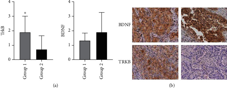
Figure 1.

Immunohistochemical detection of TrkB and BDNF protein expression in sections of primitive ADKs of the lung with (Group 1) or without (Group 2) brain metastasis. The histograms summarize the results obtained. A statistically significant increase of TrkB protein expression was observed in samples from primitive ADK in Group 1 compared with Group 2. The BDNF protein expression was higher in Group 2 in comparison with Group 1. Notice the anatomical localization of BDNF and TrkB protein expression in ADK cell. BDNF antibodies generated a prevalent nuclear dark brown immunoreaction. The TrkB antibody generated a cytoplasmic and cell membrane immunoreaction. A clear and specific TrkB and BDNF immunostaining was documented in ADKs cells in both primitive and brain metastasis in Group 1. Contrarily, in Group 2, a strong and specific BDNF immunostaining was documented in brain metastasis, but not for TrkB. The data are the mean + SD of different experiments performed in triplicate. ×20 ∗p < 0.0177.
