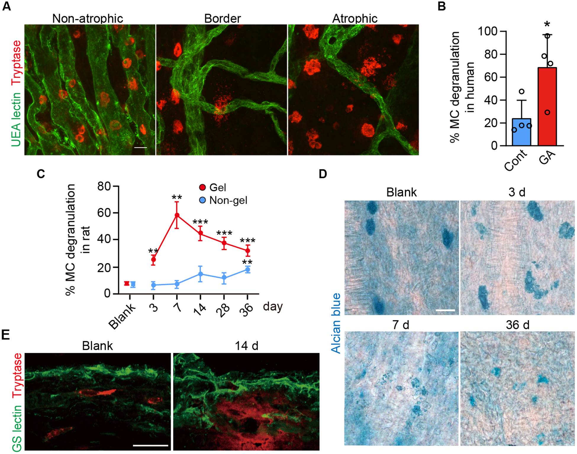
Figure 1.

Choroidal MC degranulation in human GA and in the rat model of choroidal MC degranulation. A) Choroidal flat mount immunohistochemistry showing choriocapillaris (UEA lectin; green) and MCs (anti-tryptase; red) in a human GA subject in the non-atrophic region, at the border of atrophy, and in the atrophic lesion. B) Quantification of percentage MC degranulation, as assessed by NSE staining, in the border region of the human GA subjects compared to aged control (n = 4, per group). C) The percentage of degranulated MCs over time after subconjunctival implantation of the 48/80-hydrogel in the rat on the implanted or gel region (superior choroid) and the non-gel region (inferior choroid), as assessed with Alcian blue staining. Quantification was performed on the 48/80 eyes throughout the time course and was compared to blank hydrogel eyes (without 48/80) at day 36 (blank, n = 3; 48/80, n = 4, per time point). D) Time course of MC degranulation in choroidal flat mounts stained with Alcian blue. E) Immunofluorescence of rat choroidal cross sections showing MC (tryptase+; red) and choroidal vessels (GS lectin+; green). Release of tryptase in the choroidal stroma was seen at 14 d post implantation. Data are mean ± SD. *P < 0.05, **P < 0.01, ***P < 0.001, 2-tailed, unpaired Student’s t test. Scale bars, 20 μm.
