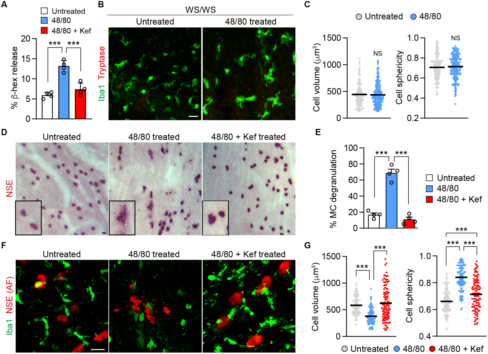
Figure 4.

Assays to evaluate MC degranulation and drug efficacy. A) In vitro microplate assay evaluating 48/80-induced (10 μg/ml) release of β-hexosaminidase from RBL-2H3 cells. Cells were co-incubated with 48/80 with or without ketotifen fumarate (Kef) (10 μg/ml) or untreated (no 48/80) medium for 45 min (n = 4). B, C) Choroidal flat mount from ex vivo eyecup assay in MC deficient WsRCws/ws (WS/WS) rats showing macrophages (Iba1+) and MC (tryptase+). Eyecups were incubated for 3 h with 48/80 (300 μg/ml). MCs were not detected in these rats. Graph showing comparison of volume and sphericity of Iba1+ cells in untreated and 48/80 treated eyes of WsRCws/ws rats (untreated, n = 165; 48/80 treated, n = 286). D) Bright field image of the choroid showing MCs (NSE+) after 3 h ex vivo in S/D rats. The boxes are higher magnification images of MCs. E) Quantification of 48/80-induced MC degranulation after 3 h, co-incubated with or without ketotifen fumarate and untreated (n = 4, per group). F) Ex vivo immunohistochemistry of S/D choroid showing MC [NSE, auto-fluorescence (AF)] at 633 nm wavelength and macrophages (Iba1+) after 3 h treatment. G) Comparison of cell volume and sphericity of Iba1+ cells treated with 48/80 with or without ketotifen fumarate and untreated (untreated, n = 114; 48/80, n = 127; 48/80 with ketotifen fumarate, n = 112). Data are mean ± SD. ***P < 0.001, 1-way ANOVA with Tukey post-hoc comparison (A, E, G), 2-tailed, unpaired Student’s t test (C). Scale bars, 20 μm.
