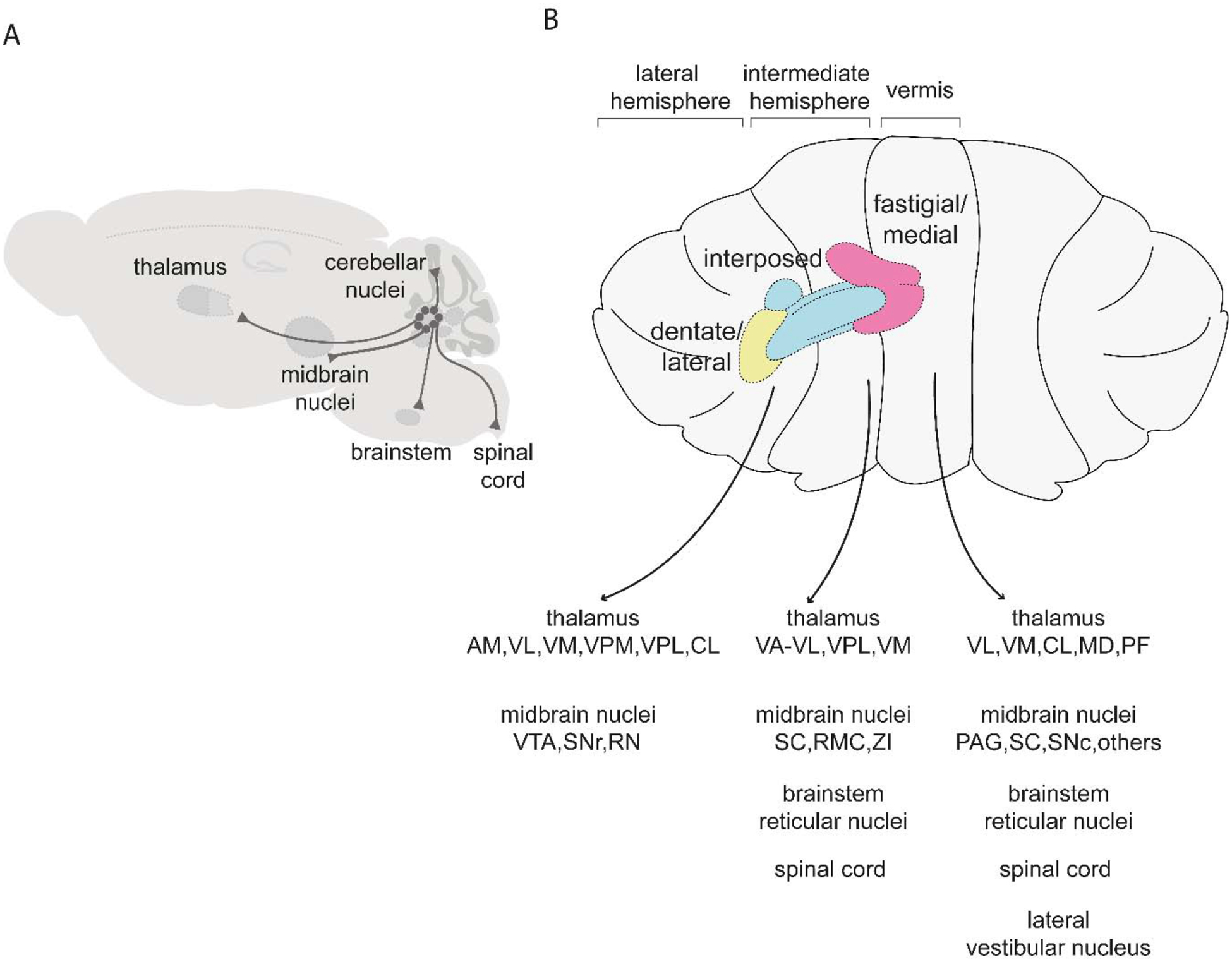
Figure 3. Major targets of the cerebellar nuclei.

(A) Schematic of pathways originating from the cerebellar nuclei. (B) Targets of the fastigial/medial nucleus include the thalamus (VL: ventrolateral, VM: ventromedial, CL: centrolateral, MD: mediodorsal, PF: parafascicular), midbrain nuclei (PAG: periaqueductal gray, SC: superior colliculus, SNc: substantia nigra pars compacta), brainstem reticular nuclei, spinal cord, lateral vestibular nucleus, and other regions described in (Fujita et al., 2020). Targets of the interposed nuclei include the thalamus (VA-VL: ventral anterior-ventrolateral, VPL: ventral posterolateral, VM: ventromedial), midbrain nuclei (SC: superior colliculus, RMC: magnocellular red nucleus, ZI: zona incerta), brainstem reticular nuclei, and spinal cord (Houck and Person, 2015; Low et al., 2018; Sathyamurthy et al., 2020). Targets of the dentate/lateral nucleus include the thalamus (AM: anteromedial, VL: ventrolateral, VM: ventromedial, VPM: ventral posteromedial: VPL ventral posterolateral, CL: centrolateral) and midbrain nuclei (VTA: ventral tegmental area, SNr: substantia nigra pars reticulata, RN: red nucleus) (Carta et al., 2019; Dacre et al., 2019; Sakayori et al., 2019). For simplicity, schematics are restricted to tracing studies performed in mice.
