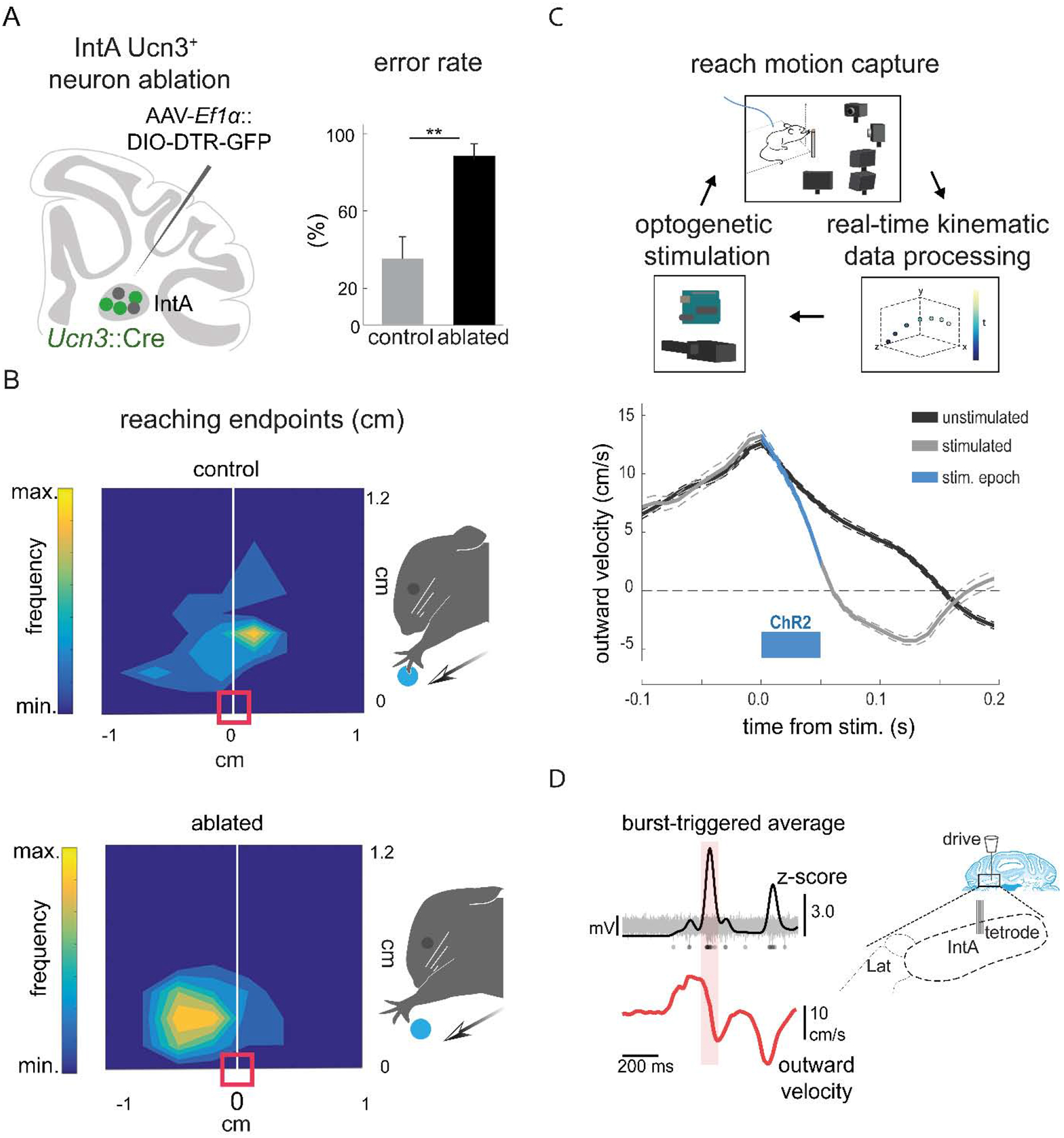
Figure 6. The IntA nucleus ensures endpoint precision during skilled reaching.

(A) Schematic summarizing strategy for selective ablation of Ucn3+ IntA neurons using a viral diphtheria toxin receptor-mediated strategy (left). Ablation of Ucn3+ IntA neurons results in increased pellet reach-to-grasp errors and reduces pellet retrieval success (right) (Low et al., 2018). (B) Contour density plots depict the endpoint position of the wrist with respect to the pellet. Arrows depict the direction of reach and the red box indicates the position of the pellet. Ablation of Ucn3+ IntA neurons results in hypermetric reaching and overshooting the target (Low et al., 2018). (C) Schematic of the behavioral setup for real-time kinematic analysis and closed-loop optogenetic manipulation of the IntA nucleus (top). Averaged velocity traces from a single animal illustrate that optogenetic activation of IntA neurons during reaching reduces outward velocity of the limb (bottom) (Becker and Person, 2019). (D) Tetrode recordings in IntA during reaching reveal that the amplitude of IntA neuronal activity (top) peaks during abrupt reductions in limb outward velocity (Becker and Person, 2019).
