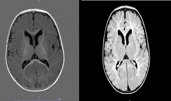
Fig. 2.
MRI of a child with Angelman syndrome at the age of 8 months. Myelination on T1w(left) shows still deficient frontal and temporal white matter and a hypoplastic corpus callosum corresponding to a maturation stage of 5–6 months. T2w image(right) indicates mildly widened ventricles, dorsally thin corpus callosum, a slightly patchy T2 signal of the white matter with particular prominence over the parieto-occipital region.

