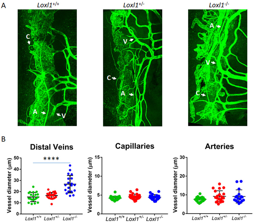
Figure 3. Loxl1−/− mice display dilated distal outflow veins but not arteries or capillaries.
(A) Loxl1−/−, Loxl1+/− and Loxl1+/+ mouse anterior segments were immunostained with antibody against CD31, and imaged using confocal microscopy at 20 × magnification. (B) Measurements of distal outflow vessels indicate a significant increase in distal vessel width in Loxl1−/− mice when compared to Loxl1+/+ eyes (****p<0.0001). There was no difference between Loxl1+/+ and Loxl1+/− eyes. The central line in the scatter plot indicates the mean (n=6 eyes from 4 mice for both Loxl1+/+ and Loxl1+/−, n=6 eyes from 5 mice for Loxl1−/−) A, arteries; V, veins; C, capillaries (scale bar=100μm).

