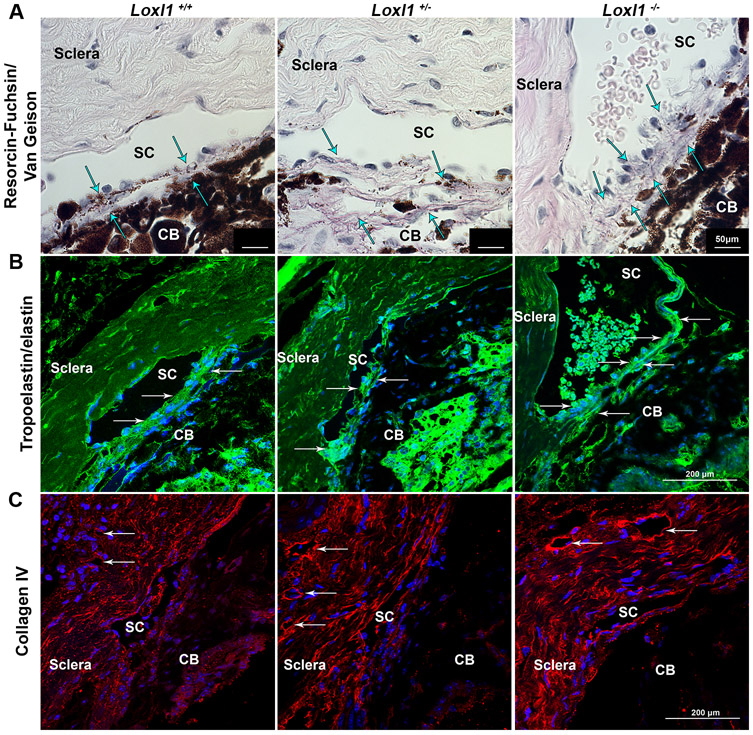
Figure 6. Loxl1−/− mice have altered distribution of collagen IV and elastin fibers in the conventional outflow pathway.
(A) Paraffin sections stained for collagen and elastin show an accumulation of elastin (arrows point to dark purple fibers) in the outflow pathway of Loxl1−/− eyes in comparison to Loxl1−/+ and Loxl1+/+ eyes, which maintained a longer fiber structure with age (n=3 mice) (scale bar = 50μm). (B) Sections from Loxl1−/− eyes indicated increased tropoelastin/elastin immunolabeling in the TM and JCT regions (arrows) in comparison to Loxl1−/+ and Loxl1+/+ eyes (n=3 mice) (scale bar=200 μm). (C) Sagittal sections from Loxl1−/− and Loxl1−/+ eyes showed increased immunolabeling of collagen IV around distal vessels (arrows) of outflow tissues in comparison to Loxl1+/+ eyes (n=3 mice) (scale bar=200 μm). Note the blood cells in the lumen of SC in the Loxl1−/− eye.

