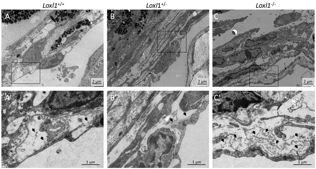
Figure 7. Loxl1−/− mice display more basal laminar deposits beneath inner wall SC endothelial cells.

Loxl1−/−, Loxl1+/− and Loxl1+/+ mouse anterior segments were embedded in Epon, sectioned, stained with uranyl acetate/lead citrate, and examined with a JEM-1400 electron microscope (JEOL USA). (A-C) Representative images from conventional outflow pathway of Loxl1−/− , Loxl1+/− and Loxl1 +/+ eyes. (A’-C’) show enlarged areas indicated by boxes in (A-C). Arrows point to basal laminar material beneath SC endothelial cells. (n=4 Loxl1+/+ mice, n=4 Loxl1+/− mice, n=5 Loxl1−/− mice).
